|
四、载药微球实验动物病理研究
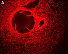
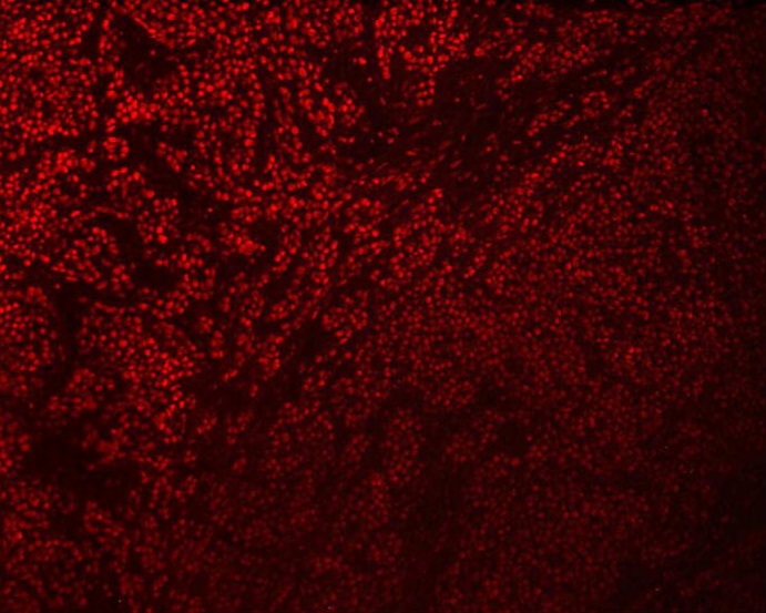
荧光显微镜(Fluorescence microscopy) 用于观察表阿霉素在肿瘤组织的分布。HepaSphere长时间持续药物释放的特性通过治疗后14天微粒周围持续性荧光存在得到进一步阐释。因为药物持续释放增加了HepaSphere导致肿瘤细胞凋亡的可能性。比起标准的TACE和肝动脉灌注,HepaSphere有较高的潜在治疗肿瘤的疗效。
Fluorescence study of spatial doxo distribution :extended 400–1600 μm into the surrounding tissue
 |
 |
|
第1天,微粒中的表阿霉素向周围组织释放 |
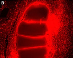
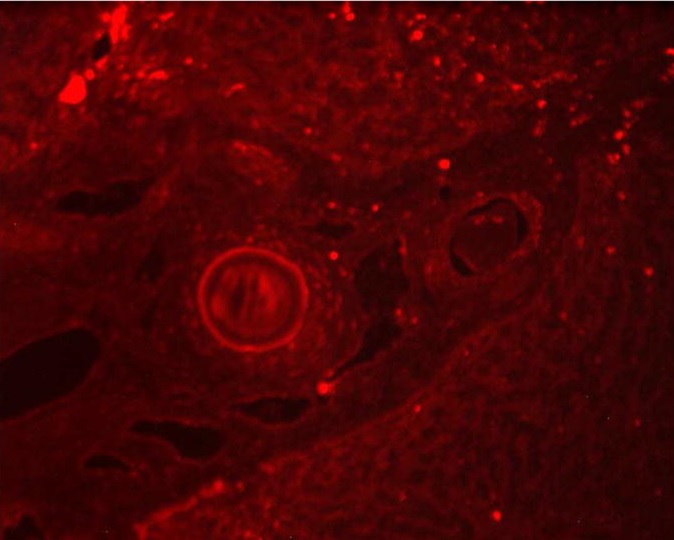
第3天,周围组织表阿霉素持续增加,微粒荧光显影渐渐变淡 |
 |
 |
|
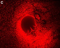
第7天,表阿霉素在周围组织持续扩大 |
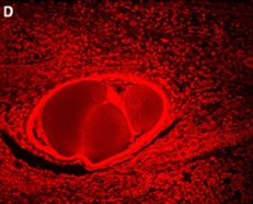
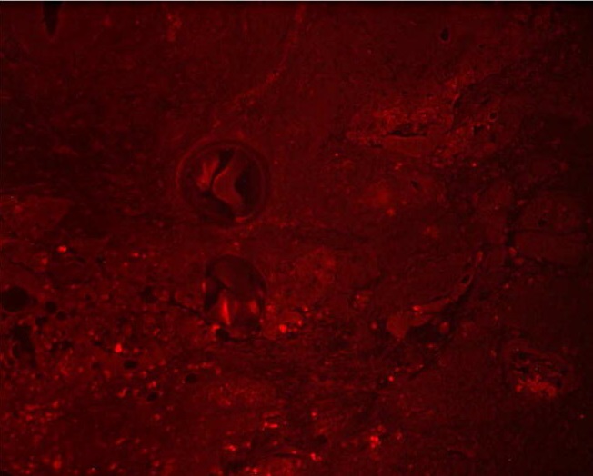
第14天,仍可以看到血管内微粒有荧光染色。 |
Gupta et al. “Hepatic Arterial Embolization with Doxorubicin-Loaded Superabsorbent Polymer Microspheres in a Rabbit Liver Tumor Model.” Cardiovasc Intervent Radiol 2011; 34(5):1021-30.
碘油-表阿霉素乳化液栓塞VX2肿瘤模型后表阿霉素荧光染色(Doxorubicin Fluorescence)
 |
 |
 |
|
c-TACE组治疗后第一天显示碘油内表阿霉素荧光随机分布遍及在肿瘤和周围正常肝组织 |
治疗后第三天,很少甚至没有表阿霉素荧光染色 |
第七天,表阿霉素的荧光染色几乎没有存在。 |
关于药物的荷载和洗脱,Gupta 等人在他们的这篇文章中讨论了表阿霉素在荧光显微镜下的表现。DEB-TACE的理由之一是随着时间的推移药物缓慢的释放。这达到了下列目标:
1. 它维持了药物在肿瘤内的长时间持续存在有助于肿瘤细胞的杀灭。
2. 药物在身体其它部位释放减少,有助于减少药物导致的副反应。
Gupta 等人的文章给人印象深刻地显示了微球如何贴服血管壁没有剩余空间的真实影像。
体外实验:多大的粒子直径合适?粒径的重要性!
动脉内注射动物模型肝癌时,为了验证是否可以用磁共振探查到不同大小的含氧化铁微球(Iron Oxide– containing Embosphere (IOE) ,和在组织学分析之前检测到微球的分布。Lee等人【3】20只新西兰白兔VX2肝肿瘤随机分组。100-300和300-500μm微导管置于肝固有动脉分别进行栓塞。
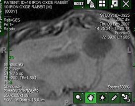
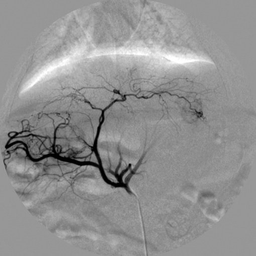
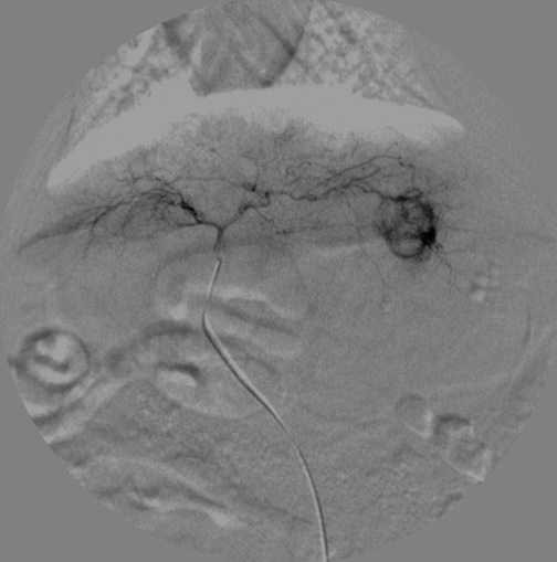
兔子肝动脉造影
 |
 |
|
腹腔动脉造影,典型的肝动脉解剖 |
左肝VX2肿瘤染色 |
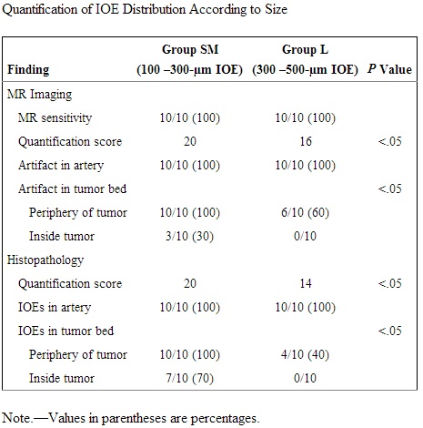
MRI检出率两组均为100%,较小粒径的顺磁性微粒100~300μm,IOE在肿瘤内检出率为30%,病例分析,IOE肿瘤内检出率为70%,大粒径300~500μm,组织学分析和MRI显示在肝动脉内的肿瘤外。
含氧化铁的微粒经肝动脉栓塞后在肝癌动物模型中的分布:使用MRI评价。
理想的药物释放: Importance of Particle Size:动脉栓塞治疗Vx-2肝脏肿瘤:含氧化铁微粒 300~500 μm 与100-300μm 比较
 |
 |
|
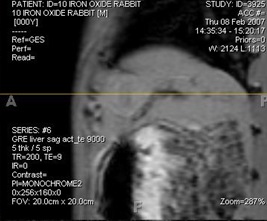
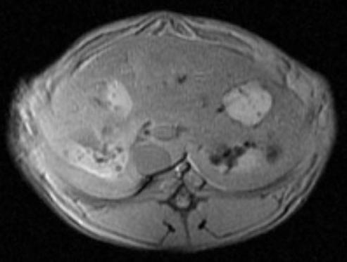
肝100–300μm IOEs 栓塞后轴位T2加权MR图像。注意点状散在信号 |
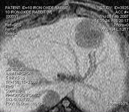
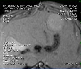
肝脏用300–500μm IOE 栓塞后的轴位T2加权MR图像。在肿瘤的边缘可见信号强度小和部分没有信号分布。肿瘤内部分没有信号 |
此实验动物表明,小粒径100~300μm被释放到肿瘤内或接近肿瘤的边缘。适用于药物释放和精确栓塞。
 |
|
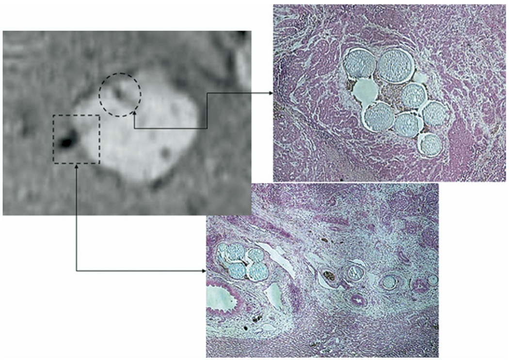
荷载氧化铁微粒100~300 μm 动脉栓塞治疗动物模型肿瘤:MR Imaging and histopathologic correlation in an animal in group S treated with 100–300-µm IOEs. Good deposition of particles in the tumor bed at the intracapsular deposition of periphery of the tumor and inside the tumor. Prussian blue ...
|
 |
|
IA Therapy for Vx-2 Liver Tumor:Iron-oxide Labeled Microspheres 100-300μm
|
为了评价栓塞后不同时间点表阿霉素在局部组织的浓度和微球内残存药物的量,以及比较动物模型中微球周围组织阿霉素的浓度水平。是否和体外药物溶出试验一样,表阿霉素洗脱微球可以维持几周的药物细胞毒性浓度。Namur等人【4】进行了试验动物研究。
药物洗脱微球的肝栓塞:表阿霉素在动物模型中的浓度
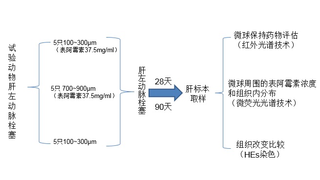
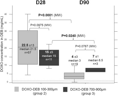
100-300μm or 700-900μm 微球荷载 37.5 mg dox/mL 栓塞后28天或90天进行肝脏标本分析,结果
1, DEBs 在28天洗脱初始药物量的43%,90天89%。药物在微球周围组织的浓度,在两个时间点上显著减少(P=0.0004)
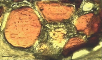
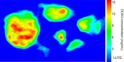
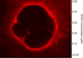
Doxorubicin quantitative mapping in DEBs
 |
 |
|
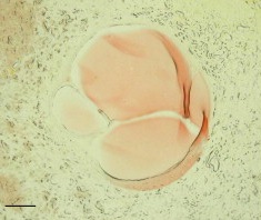
unstained tissue section of a vessel occluded by five doxorubicin DEBs (100–300 μm, day 28) |
infrared microspectroscopy image of doxorubicin inside the DEBs. Scale bar: 70 μm. |
Doxorubicin concentration in DEBs at day 28 and day 90 for the two sizes of DEBs
 |
|
Doxorubicin concentration was measured on 68 beads at day 28 and 22 beads at day 90. Dotted line indicates the LLOQ (1 mg/mL). The concentration of doxorubicin inside DEBs was not significantly different between the two sizes of doxorubicin-eluting beads at either time point. Doxorubicin concentration in the beads decreased significantly with time. |
2. 距微粒边缘600μm组织内可以检查到药物的浓度
Doxorubicin concentration profiles in the tissue around DEBs for different groups
 |
|
The tissue concentration of doxorubicin decreased with the distance from the bead for both sizes of doxorubicin DEBs. The tissue concentration of doxorubicin decreased over time in both size groups. The tissue doxorubicin concentration was higher around large DEBs than small DEBs at both time points. |
 |
 |
|
unstained tissue section of a vessel occluded by four doxorubicin DEBs (100–300 μm, day 28) |
fluorescence microspectroscopy image of free doxorubicin around the same vessel |
3. 药物在组织中的浓度范围0.55~6.8m,是肝细胞培养基内细胞毒性浓度。
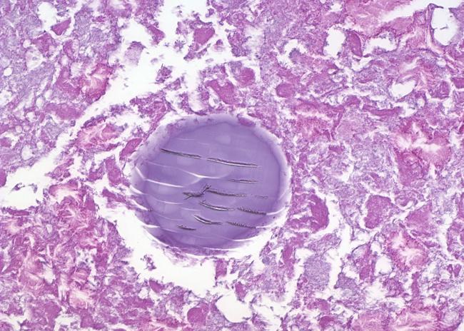
4. 药物高浓度与肝实质凝固性坏死相关联
 |
|
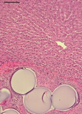
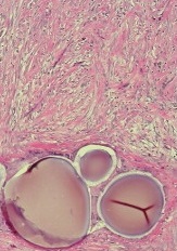
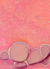
微粒周围组织坏死的范围
|
 |
 |
 |
|
normal liver parenchyma,空泡微球 |
fibrotic issue (28天)(100~300μm) |
coagulative necrosis(90天)
(100~300μm) |
5. 100-300μm 比 700-900μm 导致更多的坏死(p=0.0036),MicroCT分析:直径较小的微粒=更末梢血管=均匀的分布
6. 离微粒越远阿霉素的浓度逐渐下降,但对组织仍然有足够的细胞毒性
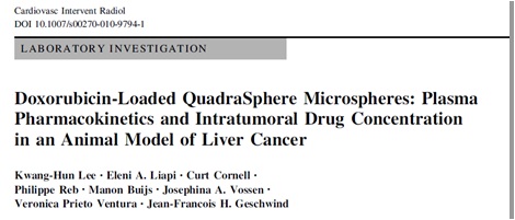
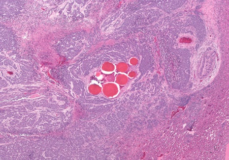
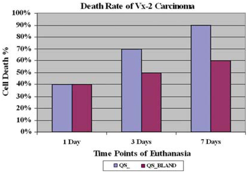
Lee等人【8】,对VX-2肿瘤实施载药微球肝动脉栓塞后进行组织学检查
载药微球与非载药微球的比较
 |
|
肿瘤坏死率的区别发生在第三天,到第七天,载药微球平均坏死率达90%,比较没有荷载药物微球的坏死率在这一天仅为60%。 |
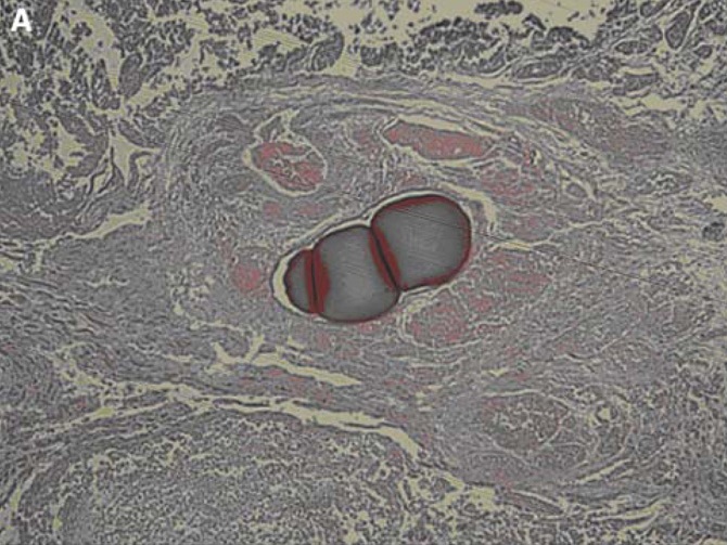
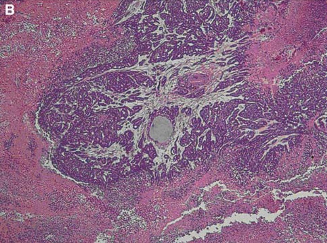
来自Lee等人【8】的同一个实验
Pathologic images obtained at 7 days after treatment according to the treatment modalities
 |
 |
|
A case treated with doxorubicin-loaded QuadraSphere microspheres shows near complete tumor cell death, whereas |
a case treated with bland embolization shows residual viable cells around the embolized area... |
(责任编辑:Mr.Editor) |