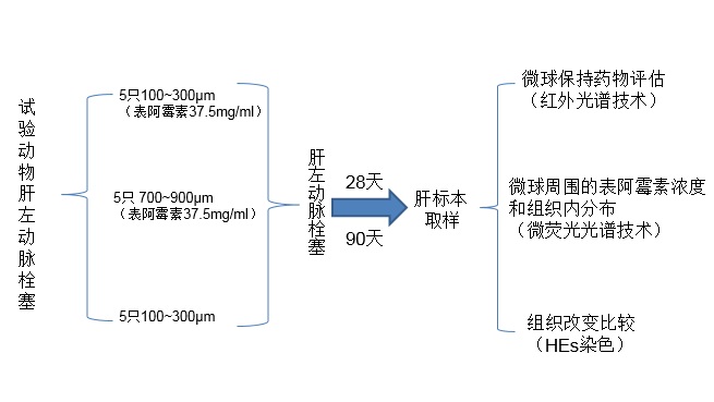
为了评价栓塞后不同时间点表阿霉素在局部组织的浓度和微球内残存药物的量,以及比较动物模型中微球周围组织阿霉素的浓度水平。是否和体外药物溶出试验一样,表阿霉素洗脱微球可以维持几周的药物细胞毒性浓度。Namur等人【4】进行了试验动物研究。
100-300μm or 700-900μm 微球荷载 37.5 mg dox/mL 栓塞后28天或90天进行肝脏标本分析,结果
1, DEBs 在28天洗脱初始药物量的43%,90天89%。药物在微球周围组织的浓度,在两个时间点上显著减少(P=0.0004)
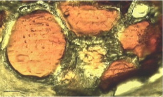
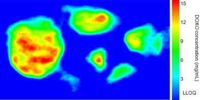
Doxorubicin quantitative mapping in DEBs
 |
 |
|
unstained tissue section of a vessel occluded by five doxorubicin DEBs (100–300 μm, day 28) |
infrared microspectroscopy image of doxorubicin inside the DEBs. Scale bar: 70 μm. |
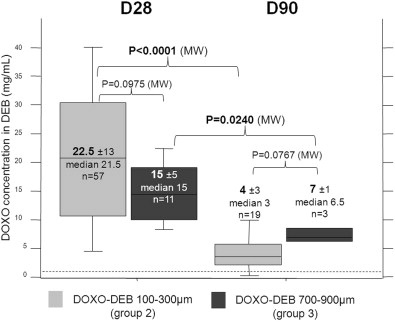
Doxorubicin concentration in DEBs at day 28 and day 90 for the two sizes of DEBs
 |
|
Doxorubicin concentration was measured on 68 beads at day 28 and 22 beads at day 90. Dotted line indicates the LLOQ (1 mg/mL). The concentration of doxorubicin inside DEBs was not significantly different between the two sizes of doxorubicin-eluting beads at either time point. Doxorubicin concentration in the beads decreased significantly with time. |
2. 距微粒边缘600μm组织内可以检查到药物的浓度
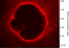
Doxorubicin concentration profiles in the tissue around DEBs for different groups
 |
|
The tissue concentration of doxorubicin decreased with the distance from the bead for both sizes of doxorubicin DEBs. The tissue concentration of doxorubicin decreased over time in both size groups. The tissue doxorubicin concentration was higher around large DEBs than small DEBs at both time points. |
 |
 |
|
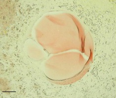
unstained tissue section of a vessel occluded by four doxorubicin DEBs (100–300 μm, day 28) |
fluorescence microspectroscopy image of free doxorubicin around the same vessel |
