目标病变的选择(在存在多个潜在目标时)
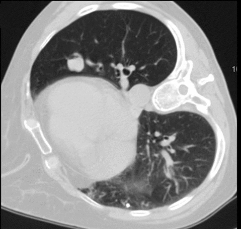
大小往往不是选择目标时最重要的因素。在一个合作的患者中,选择上肺病变,因为上肺通过呼吸移动的幅度小。然而,即使是1-2cm的病变,位于下肺即为不良病变目标,因为在最初的过胸壁、过脏层胸膜穿刺或在获得后续穿刺样本的这三个过程中的某个时刻,呼吸运动导致针尖/道与病变重合失调(misregistration)的可能性很高。
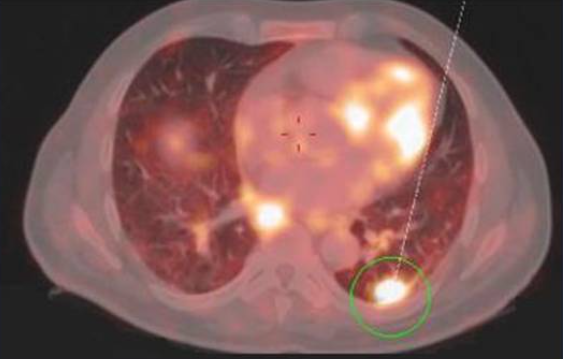
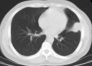
一个大的从肺门中心向肺周边延展,呈楔形或不规则形状,真正的病变以相同密度的“埋”在其中毗邻肺实质中,在视觉上并不能识别正确的病变位置,之前的PET-CT若显示有高代谢的位置,就是经皮肺病变活检的理想病变。PET/CT病变更高的代谢活动增加了获得活检的异常组织的机会。
肺外周位置的病变大大减少了穿刺大血管的机会和穿刺过程中针漂移/重定向的机会。在进入时避免肺充气的能力降低了发生气胸的几率。
肺中最容易活动的部分是横膈肌周围(下肺)。
The part of the lung most difficult to expand after a pneumothorax is the lung apex.
气胸后肺最难以复张的部分是肺尖。
为了防止或减少气胸,肺下叶上部的区域与膈肌周围病变和肺下叶尖部的病变比较,这部分的区域在患者仰卧位的时候由于受重力影响移动相对少。
In a patient with multiple lesions and prior invasive thoracic procedure, the side on which prior surgery was performed should be selected for biopsy.
对于有多发性病变且既往有侵入性胸部手术的患者,应选择既往进行过手术的一侧进行活检。
Pleural adherence can increase due to scarring and reduce the chance of a pneumothorax.
胸膜粘附可因瘢痕形成而增加,并减少发生气胸的机会。
Puncturing via the thoracotomy scar itself should be avoided, however, because the hard tissue can interfere with needle manipulation.
然而,应该避免通过开胸疤痕本身进行穿刺,因为硬组织可能会干扰针的操作。
It cannot be stressed enough that the morphology of the area of concern is a key feature when contemplating lung biopsy.
需要强调的是,在考虑肺活检时,所关注区域的形态是一个关键特征。
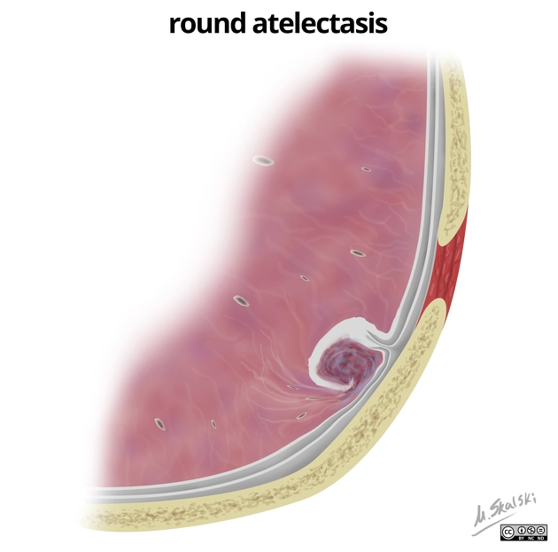
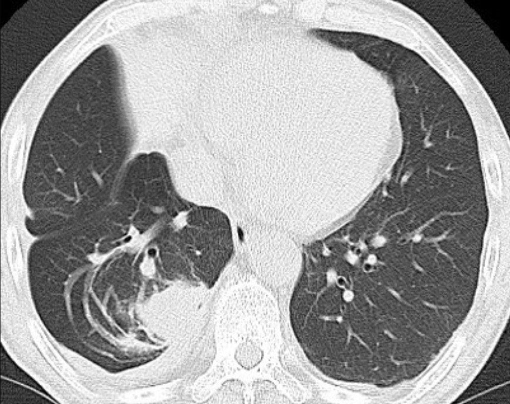
Benign round atelectasis or even "standard non-round atelectasis" can fit of all of the criteria of the optimal "lesion" as described above, including that of being very hypermetabolic on PET, as could a region of parenchymal fibrosis.
良性圆形肺不张或甚至标准非圆形肺不张,影像上可符合所有理想活检病变标准,包括PET代谢也可能非常高。
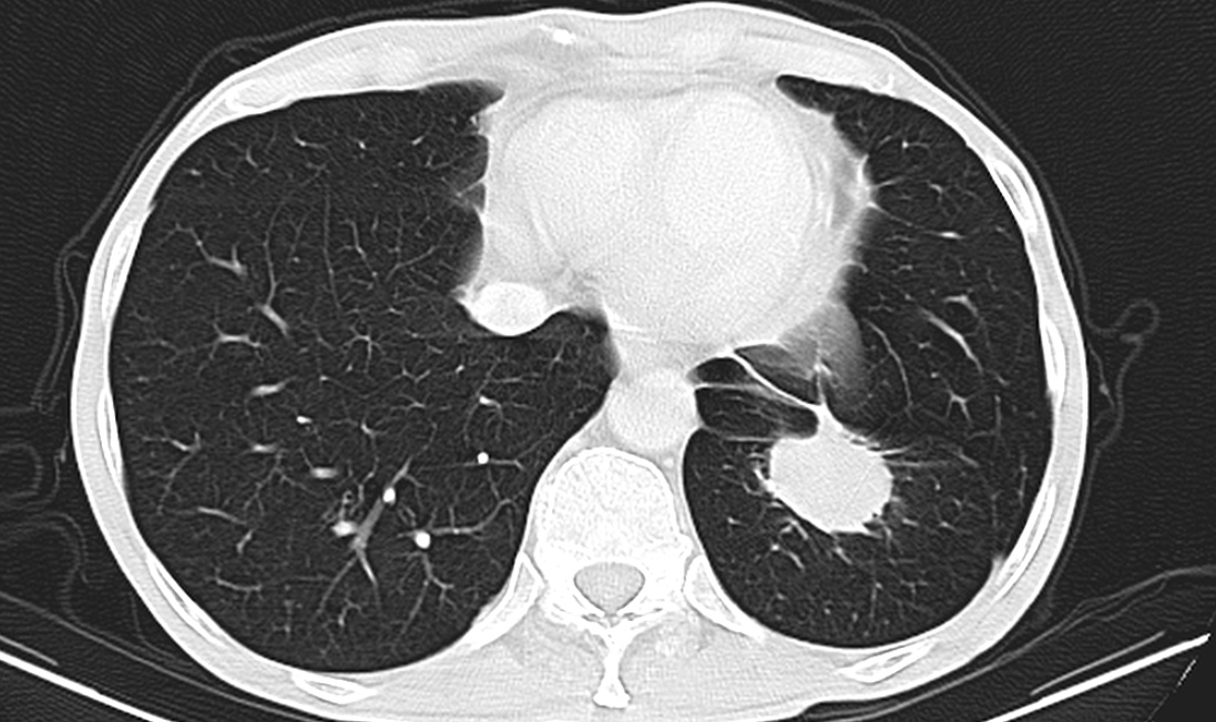
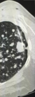
良性圆形肺不张是肺不张的不常见类型,是多余胸膜包裹形成的肺内病变。其发生的相关因素包括石棉肺、气胸治疗后,充血性心力衰竭,肺炎旁渗出等。主要表现为圆形的实变肿块。支气管内空气影可出现在肿块内。病灶多与胸膜异常相邻,包括胸膜增厚,胸膜钙化。血管和支气管卷曲进入肿块被称为“彗星尾征”(comet tail sign)。受累肺叶容积减少与肿块大小成比例。
除了形态各异,几乎有胸膜的地方都可能出现球形肺不张
However, biopsying such abnormalities either with or without an actually associated mass almost never yields clinical benefit.
然而,这种病变活检,无论有无实际相关的肿块,几乎都不会产生临床效益。
Only true nodules/masses are likely to yield any useful material on pathologic review.
只有真正的结节/肿块才有可能产生任何有用的病理检查材料。
在有些情况下,活检失败的可能性(无论是否需要紧急手术)比不失败的可能性更大(风险大于可能产生的好处)
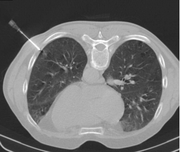
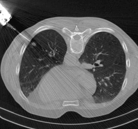
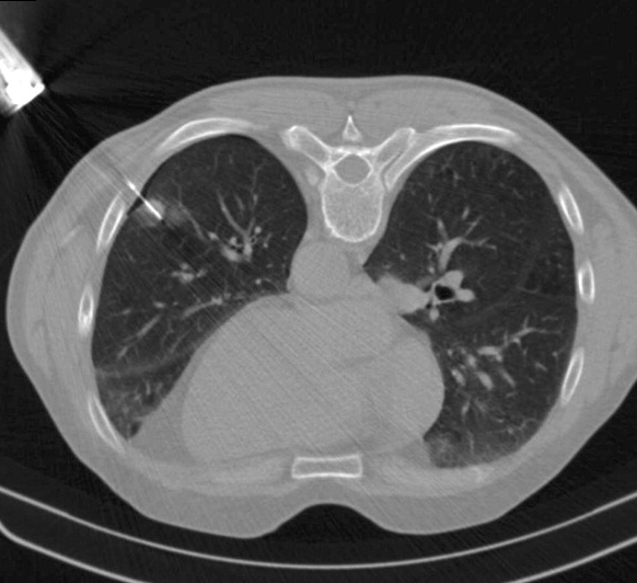
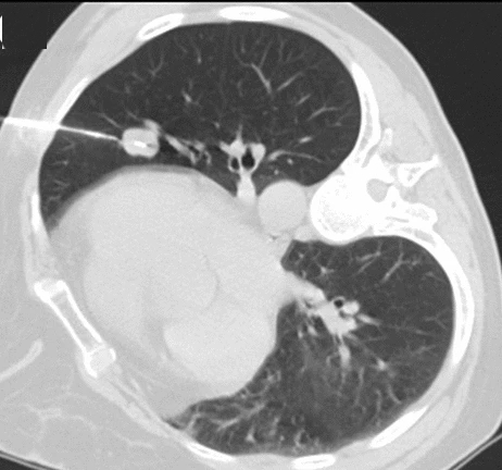
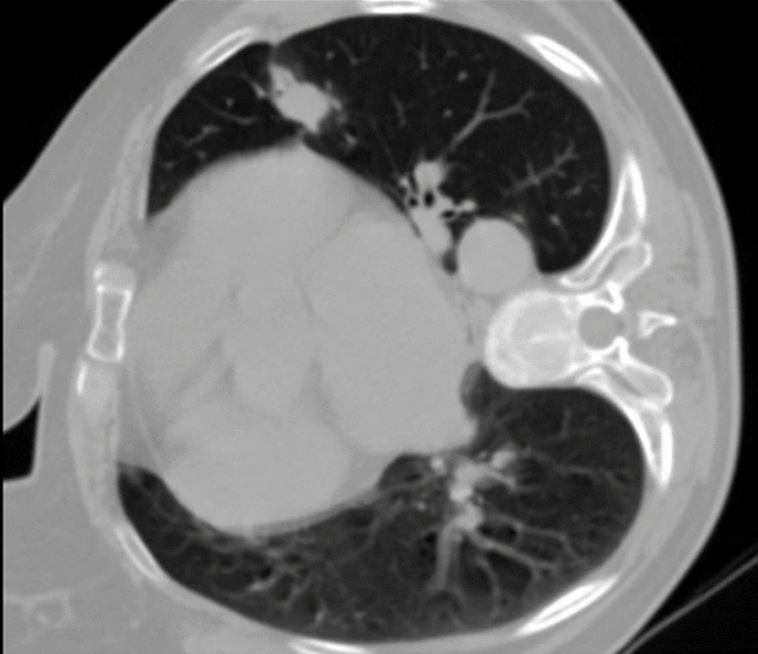
Small immediately subpleural lesions make it very difficult to time needle entry so as to pierce these lesions on the first attempt and to keep the introducer needle in the lesion over the course of obtaining multiple samples (increasing risk for pneumothorax).
小的紧贴胸膜下病变很难一次穿刺成功,而且在直接穿刺获得多个样本的过程中将引导针保持在病变中,除了增加穿刺针脱靶几率也增加了气胸的风险。采用经过更多肺组织的斜向穿刺入路可能增加获取有效样本的几率,减少气胸风险。
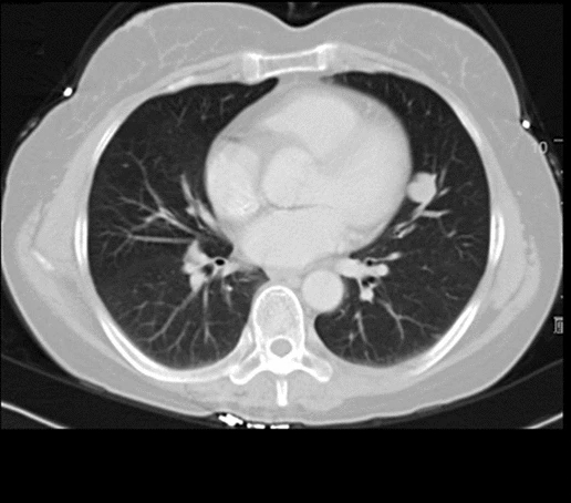
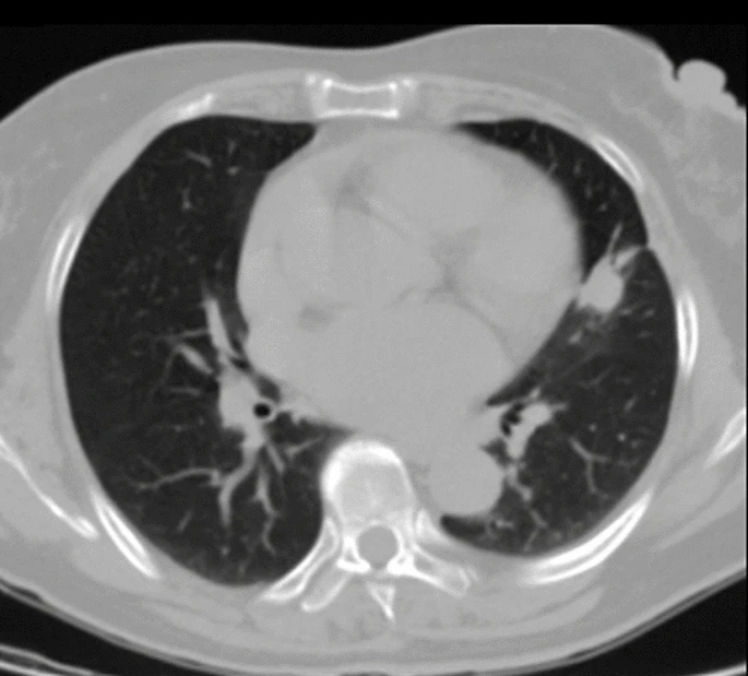
Small lesions touching the heart are usually unsuitable for biopsy.
接触心脏的小病变通常不适合活检。特别是对于初学者(beginning learners)
Cardiac motion on the lesion increases the risk of missing the lesion while placing the patient at high risk for pericardial/myocardial puncture followed by tamponade and death.
心脏运动增加了穿刺时穿刺针丢失病变的风险,同时使患者处于心包/心肌穿刺后心包填塞和死亡的高风险之中。
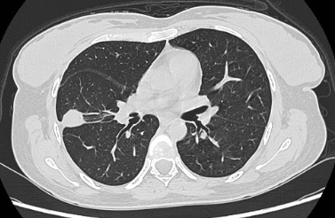
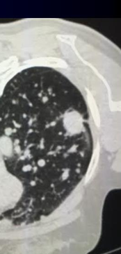
Nodules along fissures (particularly in the superior portion of the right middle lobe just under the minor dome-shaped fissure) can be difficult to target without puncturing a second lobe (thereby making two additional pleural punctures inadvertently) even with the benefit of an angled CT gantry.
沿叶间裂的肺结节(特别是在右中叶的上部,就在小的穹顶裂下),即使有角度的CT门架,也很难在没有穿刺第二叶的目标(从而无意中造成两次额外的胸膜穿刺)。
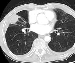
沿肺叶间裂生长的肺结节
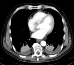
Small lesions deep in the costophrenic sulci move a great deal.
肋膈角深处的小病变移动大量。
Inadvertent puncture of the diaphragm (which can be very painful even if there is not associated puncture of an immediately underlying structure such as liver or spleen) is a likely sequela.
无意中穿刺横膈膜(这可能是非常痛苦的,即使没有相关的穿刺一个直接的基础结构,如肝脏或脾脏)是一个可能的后遗症。
Ground glass or largely necrotic lesions where the viable cellular yield is likely to be too low from needle sampling to enable a diagnosis.
磨玻璃或大部分坏死病变可能的癌细胞数量太小,以至于活检仍无法诊断。
The smaller the lesion, the more unlikely it is to yield diagnostic material.
病变越小,就越不可能产生有效诊断取样。
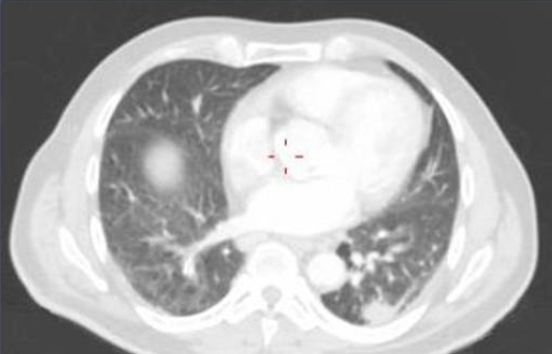
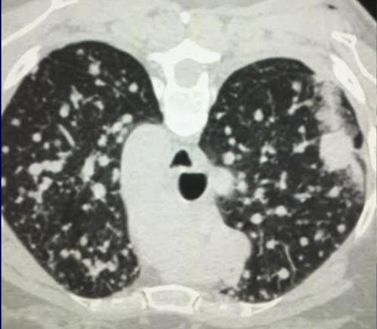
Haematogenic secondary spread of a primary lung tumor (adenocarcinoma)。Lesions should be punctured peripherically targeting it’s wall.
Nodules as small as 4 mm have yielded enough tissue for a pathologist to feel confident to diagnose nonsmall cell lung cancer [7], but size in and of itself is not a reliable indicator of success.
小至4mm的结节已经产生了足够的组织,使病理学家有信心诊断非小细胞肺癌【Li 1996】,但其大小本身并不是成功的可靠指标。
Molecular testing agencies may require at least 100 viable tumor cells (author's experience), and no study has been published that equates a number of viable tumor cells to lesion size.
分子检测机构可能需要至少100个活的肿瘤细胞(作者的经验),而且还没有发表过将一些活的肿瘤细胞等同于病变大小的研究。
In these situations, either planning to offer thoracoscopic biopsy post-percutaneous lung lesion biopsy the same day or next day service or proceeding straight to thoracoscopic sampling with all its inherent risks is the proper route.
在这些情况下,无论是计划在当天或第二天提供胸腔镜肺病变活检,还是直接进行具有所有固有风险的胸腔镜取样,都是正确的途径。
| |||||||||||||||||||||||||||||||||||||||


