Approach route
Transfemoral
Transjugular
Efferent vein embolization
Intra-procedural Cone-Beam CT protocol
The role of CBCT in determining technical success
-
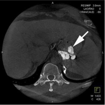
CBCT provides endpoint of gelfoam injection
√ Easily difine extent of embolization
√ Identifying nonfilling varices and veins
-
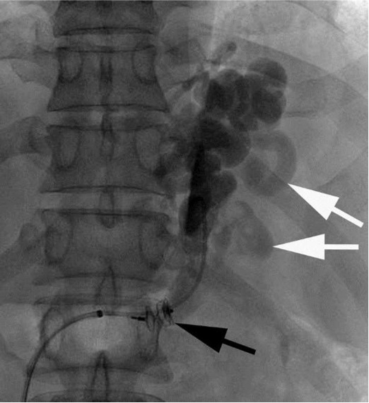
Visualization of incomplete embolization on CBCT in two patients
√ Increased technical success rate
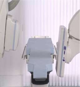 |
A flat-panel detector angiographic system
(AXIOM Artis Zee; Siemens, Erlangen, Germany)
Scan time: 7 sec
Total scanning angle, 200°
Rotation speed, 30°/s
Total of 396 projections (30 frames/sec)
Matrix size, 512 x 512
Isotropic voxel size, 0.49 mm
Effective field of view, 382 x 296 mm2
|
|
|
|
 |
 |
|
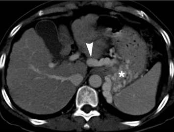
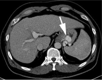
contrast CT images:afferent vein |
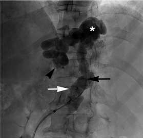
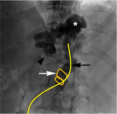
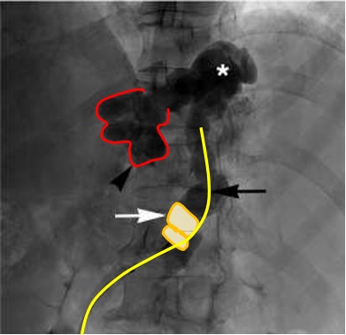
Proximal part of afferent vein was identified on the fluoroscopy |
JVIR, 2015, Gwon DI et al
 |
 |
|
JVIR, 2015, Gwon DI et al. |
|
 |
 |
|
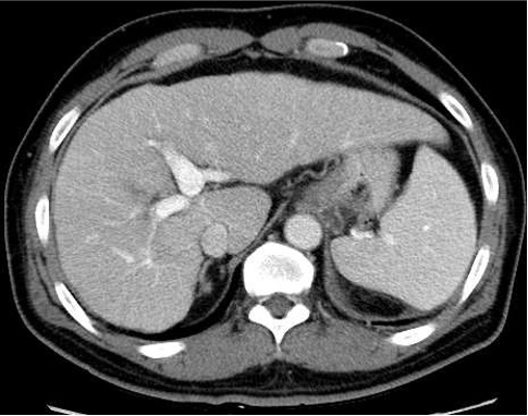
Before PARTO |
CBCT provides endpoint of gelfoam injectio
√ Easily difine extent of embolization
√ Identifying nonfilling varices and veins
|
 |
|
10mm AVP II |
 |
 |
|
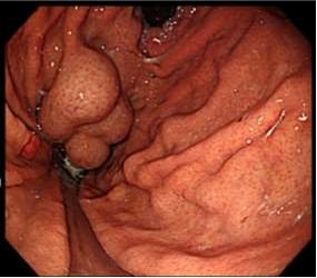
Before PARTO |
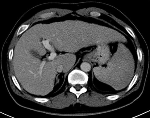
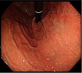
After PARTO |
 |
 |
|
Before PARTO |
After PARTO |
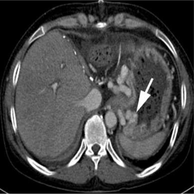
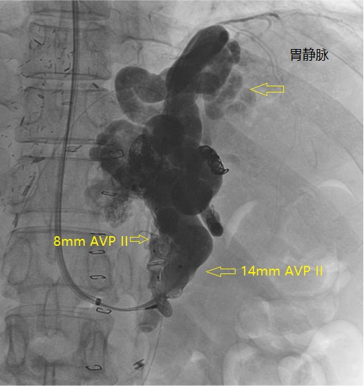
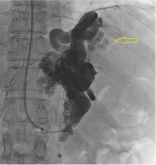
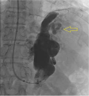
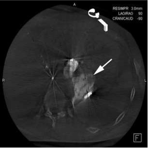
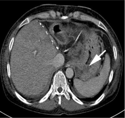
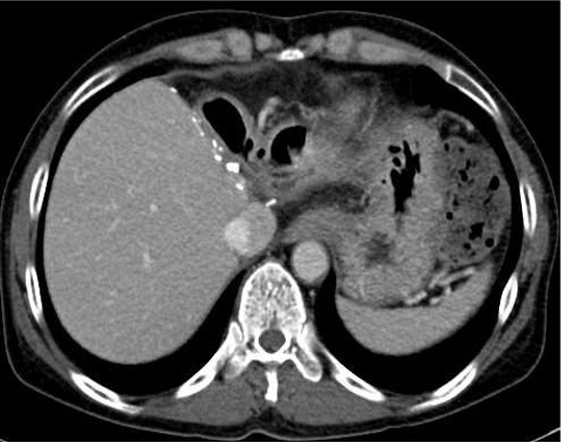
CASE 2. A 48-yr woman with two dominant gastrorenal shunt with GV s/p LT d/t alcoholic LC
 |
 |
|
Before PARTO |
1st embolization |
 |
 |
|
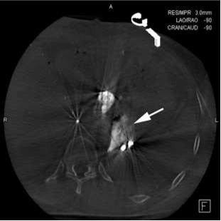
1st embolization:incomlete thrombosis of GV |
1st CBCT |
 |
 |
|
2nd embolization |
2nd CBCT |
 |
 |
|
1 day after PARTO |
4 Mo after PARTO |
PARTO is technically simple and safe and is clinically effective for the treatment of GV and HE.
Intra-procedural CBCT can be considered as an adjunct tool to fluoroscopy during PARTO.
|


