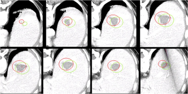
在肿瘤介入治疗成像中首要的角色是操作规划的成像。对于这个角色,首先需要的是高质量的诊断影像。在很多情况下,需要多种影像工具进行评价。有些是解剖研究,如CT或MRI影像,有些研究却是生理研究如PET或SPECT,它们可以证实肿瘤代谢方面的情况。但是,以前检查的结果也可有助于疾病活跃性的研究,应该与现在的检查进行比较以评价疾病的进展和决定计划干预的医学适宜性。 无论介入治疗是否是肿瘤消融或经导管栓塞,操作前病人评估的影像学基本内容包括 1. 介入治疗是否是医学适应症吗? 2. 介入治疗是否在技术上可行? 3. 治疗目标的最佳方法是什么? 4. 有解剖的变异吗? 5. 对邻近结构的潜在有害的影响是什么? 目前,复杂的治疗计划系统应用于影像引导下放疗和近距离治疗优化了放射治疗的效果。而类似的肿瘤介入治疗系统尚在起步阶段,处在初始开发当中。肿瘤介入治疗计划系统的一个例子是个提供涵盖以前消融病灶的消融图像。这一计划可以确保有足够的肿瘤覆盖和避免损伤重要解剖。数学模型被用来预测完全肿瘤治疗所需要的消融体积【10-12】。这些模型考虑了所需要的消融容积并然后规定包含整个肿瘤所需要的重叠消融次数。在可行性研究中,机器人甚至已经被用于执行这个重叠的消融计划【9】。影响引导用于计划射频消融电极针位置特别重要,特别是当放置多极针时候。这一重要性不仅是确保整个肿瘤得到治疗,也包括治疗的优化和安全。 Treatment planning for thermal ablation
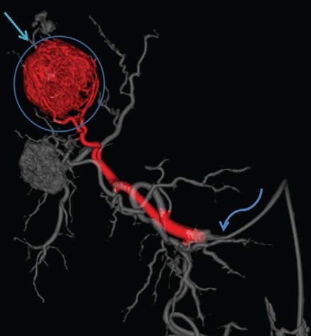
肿瘤介入计划的其它例子包括血管内肿瘤治疗,例如肝动脉栓塞和放射性微粒栓塞。肝肿瘤TACE前CT血管造影可以提供关于肿瘤滋养血管和学挂解剖变异的重要信息【13】。例如栓塞前CT血管造影可以显示右膈下动脉残余肝癌的供血【14】。内照射前(Radioembolization)多影像模式可以用于发现潜在操作并发症,例如发现来自于肝动脉的肝外血流【15】,而鍀99影像能够用于知道剂量和如果肝外发现鍀99-MAA吸收就可以避免靶标用药【16】。 有时术前计划成像可以术前就开始,但必须依赖操作中的影像完成这个计划。例如,病人介入操作的位置可以不同于术前的影像。就需要在另外的鍀病人新位置的术前计划影像。器官的位置(例如肠道)经常会移动,需要知道操作时的位置,因此需要另外的影像计划手术的实施。其它需要在操作室的计划影像包括高空间分辨率的C臂CT,其经常用于在操作时补充术前,较高对比度分辨率而较低空间分辨率的术前CT引导治疗计划成像。 在血管造影期间,旋转C臂CT提供高空间分辨率(但低对比度分辨率)类CT图像。这些三维血管造影可以分段提供术中血管内治疗计划【17】。
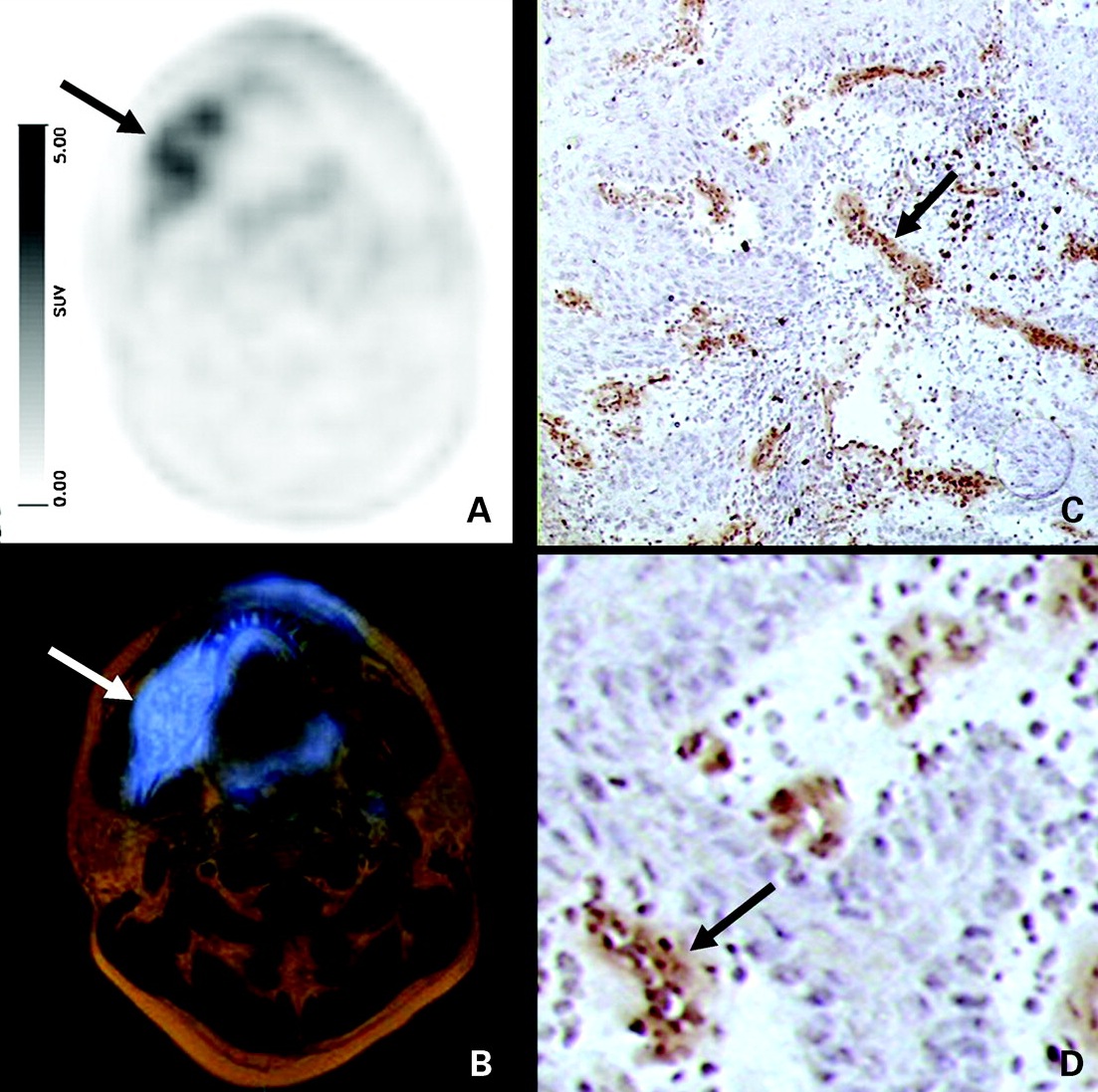
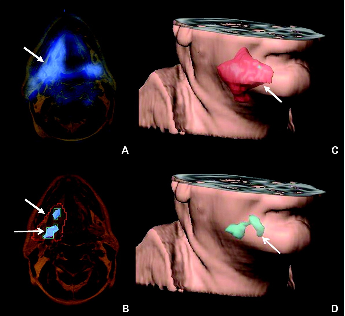
分子影像技术在临床应用的增加并将可能应用于计划和引导肿瘤介入治疗【18】。细胞增殖现象( cell proliferation imaging)【19】、血管生成显像(angiogenesis imaging)【20】和磁共振波谱分析(MR spectroscopy)【21】可以用于肿瘤介入的治疗计划成像,并可能激发新型介入靶向治疗。
Simulation systems are also being developed that allow interventional oncologists to both review images for planning purposes and to work with instruments in a virtual procedure. Recently, many institutions are applying simulation software and devices to allow physicians the opportunity to “practice” prior to the actual procedure ( 22 , 23 ). While most of these systems are still nascent, it is likely that simulation will play an increasing role in the education and training of interventional oncologists and in planning of interventional procedures in the future
9.Solomon SB , Patriciu A , Stoianovici DS . Tumor ablation treatment planning coupled to robotic implementation: a feasibility study .
( 24 , 25 ).
仿真系统也正在被开发,允许肿瘤介入医生审查术前计划影像和与操作器械一起工作的虚拟的程序。许多机构正在应用模拟软件和器械,让医生在实际操作之前,有“实践”机会【22,23】。虽然大多数的这些系统仍处于初期阶段,它是可能在将在肿瘤介入医生的教育、培训和术前计划成像中发挥越来越大的作用【24、25】。
J Vasc Interv Radiol 2006 ; 17 ( 5 ): 903 – 907 .
10. Khajanchee YS , Streeter D , Swanstrom LL , Hansen PD . A mathematical model for preoperative planning of radiofrequency
ablation of hepatic tumors . Surg Endosc 2004 ; 18 ( 4 ): 696 – 701 .
11. Jankun M , Kelly TJ , Zaim A , et al . Computer model for cryosurgery of the prostate . Comput Aided Surg 1999 ; 4 ( 4 ): 193 – 199 . 12. Villard C , Baegert C , Schreck P , Soler L , Gangi A . Optimal trajectories computation within regions of interest for hepatic RFA
planning . Med Image Comput Comput Assist Interv 2005 ; 8 ( pt 2 ): 49 – 56 .
13. Sze DY , Razavi MK , So SK , Jeffrey RB Jr. Impact of multidetector CT hepatic arteriography on the planning of chemoembolization
treatment of hepatocellular carcinoma . AJR Am J Roentgenol 2001 ; 177 ( 6 ): 1339 – 1345 .
14. Basile A , Tsetis D , Montineri A , et al . MDCT anatomic assessment of right inferior phrenic artery origin related to potential supply to hepatocellular carcinoma and its embolization . Cardiovasc Intervent Radiol 2008 ; 31 ( 2 ): 349 – 358 . 18, Torigian DA , Huang SS , Houseni M , Alavi A . Functional imaging of cancer with emphasis on molecular techniques . Cancer 2007 ;
57 ( 4 ): 206 – 224 .
19. Yang YJ , Ryu JS , Kim SY , et al . Use of 3 9 -deoxy-3 9 -[18F]fluorothymidine PET to monitor early responses to radiation therapy in
murine SCCVII tumors . Eur J Nucl Med Mol Imaging 2006 ; 33 ( 4 ): 412 – 419 .
20. Beer AJ , Grosu AL , Carlsen J , et al . [18F] galacto-RGD positron emission tomography for imaging of alphavbeta3 expression on
the neovasculature in patients with squamous cell carcinoma of the head and neck . Clin Cancer Res 2007 ; 13 ( 22 pt 1 ): 6610 – 6616 .
21. Arias-Mendoza F , Smith MR , Brown TR . Predicting treatment response in non-Hodgkin’s lymphoma from the pretreatment tumor content of phosphoethanolamine plus phosphocholine . Acad Radiol 2004 ; 11 ( 4 ): 368 – 376 . 22. Villard C , Soler L , Gangi A . Radiofrequency ablation of hepatic tumors: simulation, planning, and contribution of virtual reality and haptics . Comput Methods Biomech Biomed Engin 2005 ; 8 ( 4 ): 215 – 227 . 23. Villard PF , Vidal FP , Hunt C , et al . A prototype percutaneous transhepatic cholangiography training simulator with real-time breathing motion. Int J CARS 2009 ; 4 ( 6 ): 571 – 578 . 24. Dawson S . Procedural simulation: a primer . J Vasc Interv Radiol 2006 ; 17 ( 2 pt 1 ): 205 – 213 . 25. Lendvay TS , Hsieh FJ , Hannaford B , Rosen J . The biomechanics of percutaneous needle insertion . Stud Health Technol Inform 2008 ;132 : 245 – 247 . |


