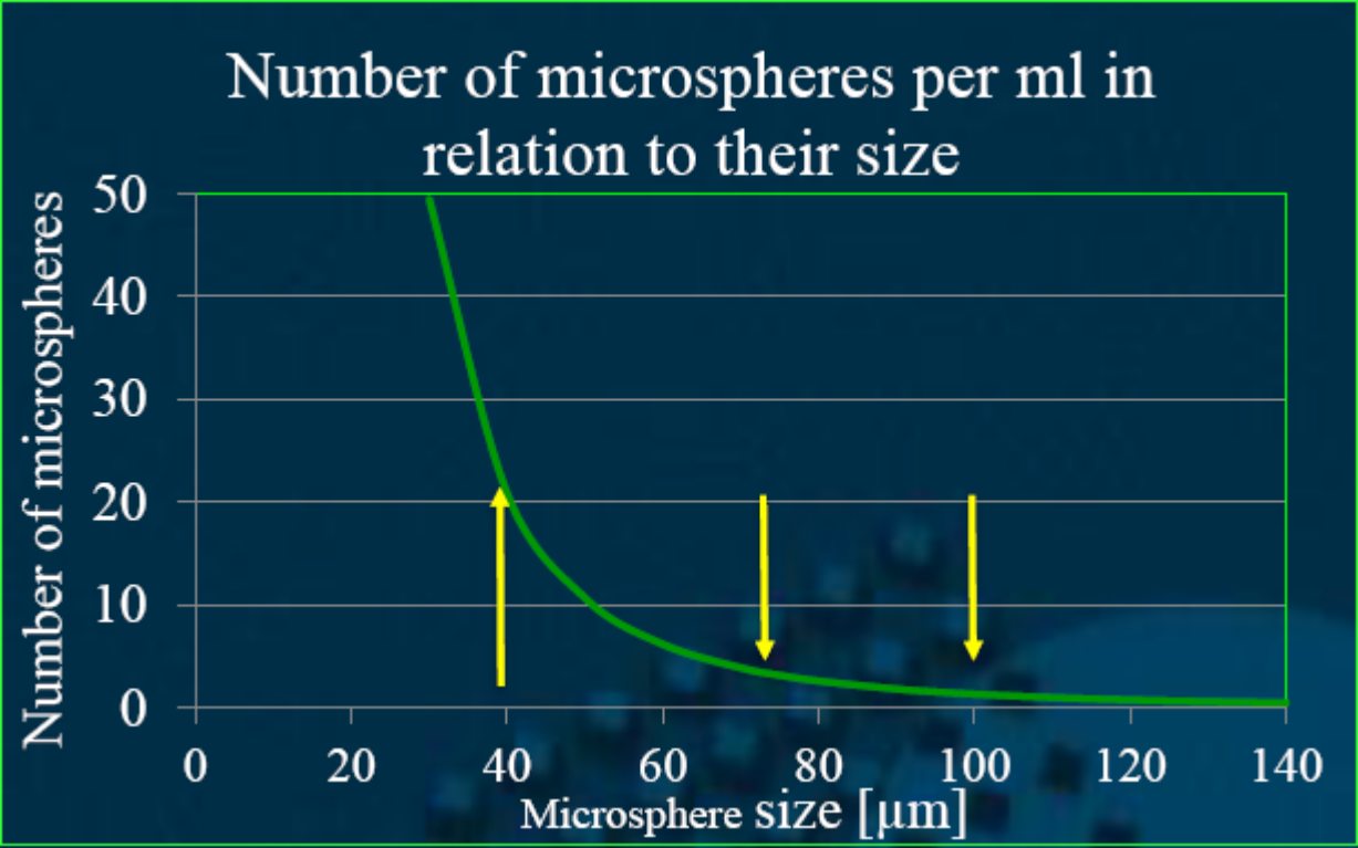
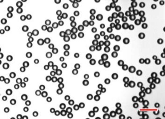
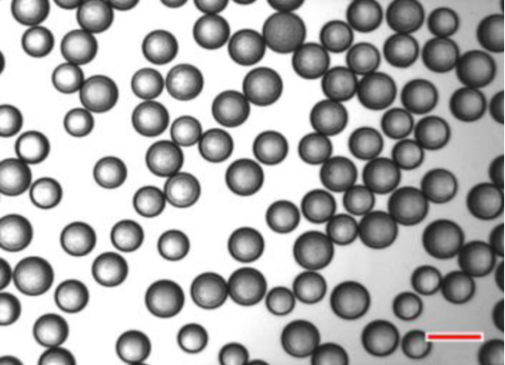
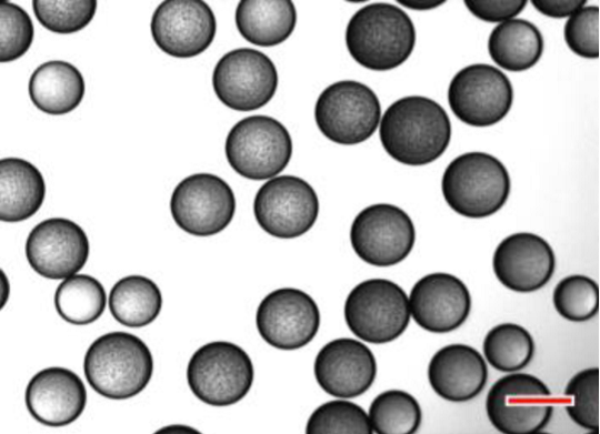
小粒径栓塞剂的定义 小粒径没有明确的定义,一般指40-120μm粒径的球形栓塞微粒。为什么需要小粒径? 栓塞关粒径什么事(Size matters)?
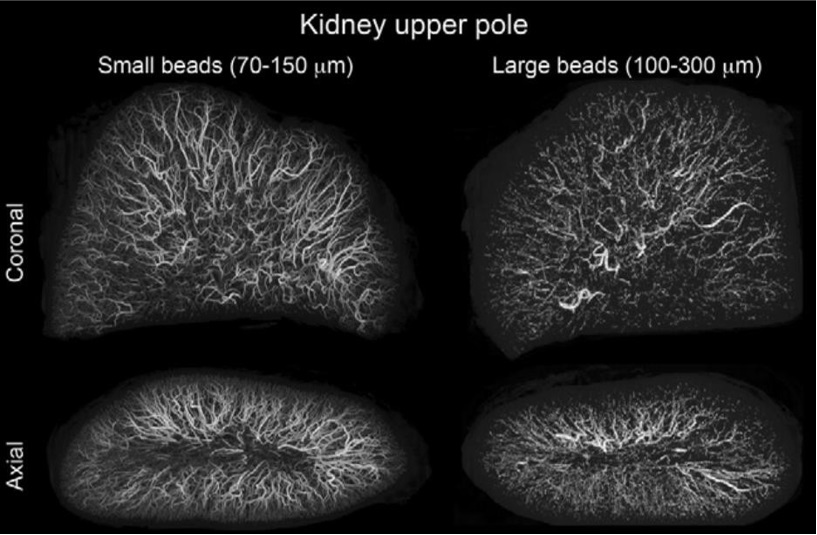
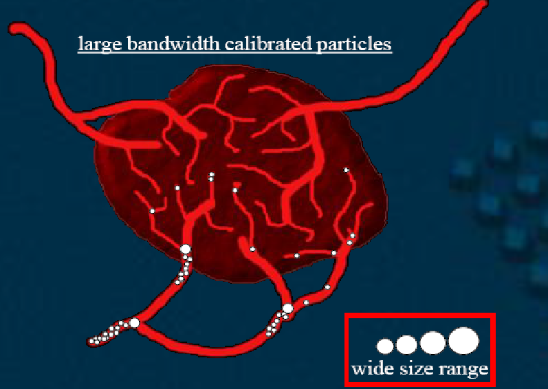
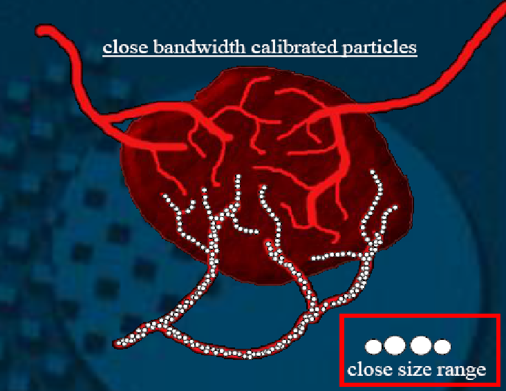
减少微粒的大小的主要原理是粒径越小,更远的(更末梢)的栓塞 Rationale: smaller bead size → more distal embolization
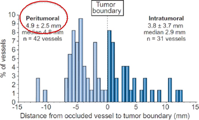
100-300𝜇 DEB block vessels with a mean diameter of 237μm
42% of vessels occluded inside the tumor; the mean distance of bead penetration inside the tumor was 3.8 ± 3.7 mm 42%的微球在肿瘤内
58% of occluded vessels outside the tumor, at a mean distance of 4.9 ± 2.5 mm from tumor boundary(58%的微球在肿瘤外)
减少微粒的大小的另一个原因 除了栓塞更远端血管而且分布也最均匀 The smaller the more distal and better spatial resolution
“Histologic evaluation of resected livers suggests that only half the beads reside in a targeted tumor versus normal liver”(J Hepatol 2011)
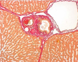
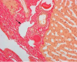
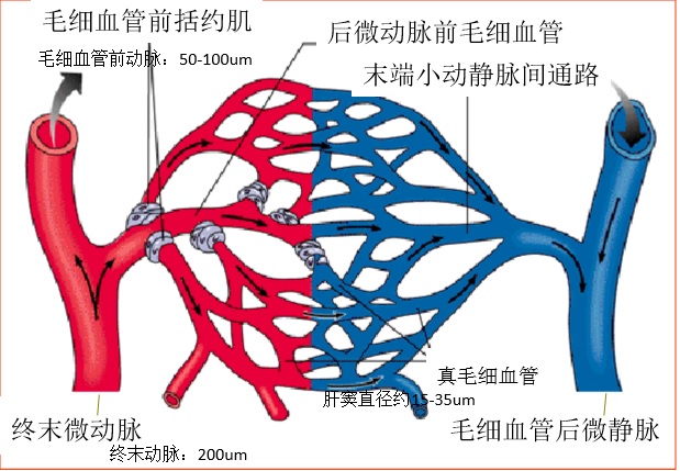
小粒径栓塞微球栓塞平面更低,甚至可达到肝窦前血管,且安全和非劣效。40-120um的粒子可以靶向性地到达肝窦前血管(50-100um),周围肝实质和胆管未见损伤
Stampfl S, Stampfl U, Rehnitz C et al (2007) Experimental evaluation of early and long-term effects of microparticle embolization in two different mini-pig models. Part II: liver. Cardiovasc Interv Radiol 30:462–468
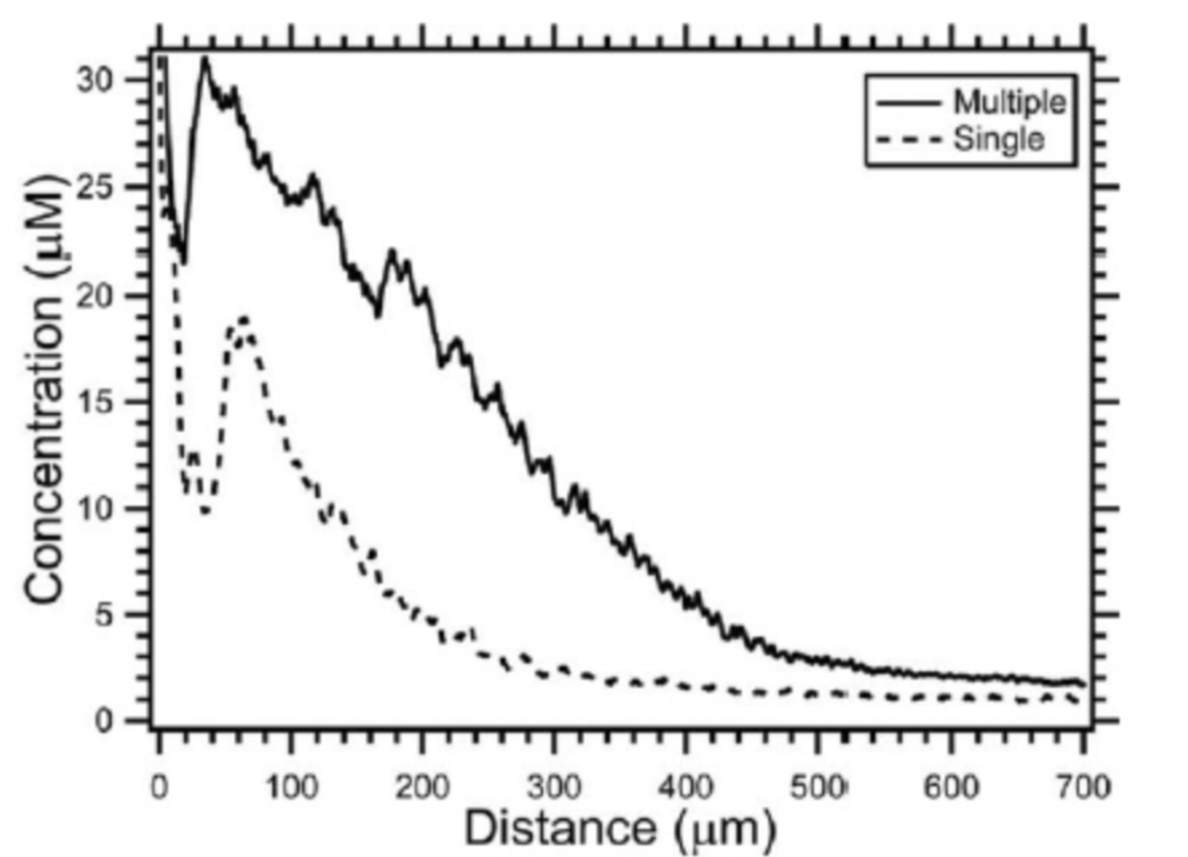
假设小粒径Quadrasphere(QS)可以释放表阿霉素到肿瘤内更为末梢的地方,并改善药物的覆盖。相反大粒径QS荷载药物的量较大,可以提供更长时间的释放。D‘inca等人比较30-60μm与50-100μm(干粒径)两种不同粒径的QS在猪模型的研究结果,比较坏死范围,QS的再分布和表阿霉素在血浆、组织和微粒的浓度水平。 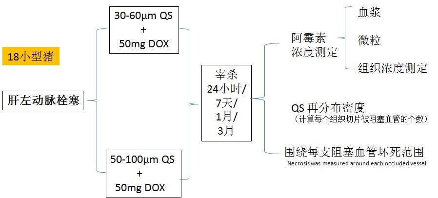 1. 第七天小粒径组的坏死范围明显大于大粒径组(P=0.0429) 2. 第1和3月没有坏死 3. 表阿霉素血浆浓度没有差异 从QS释放率
QS周围表阿霉素的浓度在第七天为382μmol/g(30-60μm组)和1138μmol/g(50-100μm组),p<0.0001,以后并没有什么不同。QS阻塞血管的数目,小粒径组明显多于大粒径组(8.0 vs 3.6,p=0.015)。 结论:用小粒径栓塞,肝坏死范围大于大粒径组。这一效果并不能被表阿霉素的释放率所解释(小粒径较快),或被组织浓度所解释(大粒径浓度较高),但和被栓塞组织内的较高的QS密度有关。即粒径越小,组织内粒子越多 除了粒径小之外,粒径均匀也非常重要
•临床前研究表明:
- 更小的微球穿透血管更远,空间密度更高【Dreher MR, et al. 2012;Lee KH, et al. 2008; Tanaka T, et al. 2014; Pelage JP, et al. 2002;】
- 均匀大小的颗粒即缩窄栓塞颗粒的大小范围,与允许控制栓塞 (即颗粒的大小与动脉闭塞的近端-远端水平相关)【Pelage JP, et al. 2002;】
• 更小的微球可获得更大的空间频率,可提供更大的药物覆盖,也就是说小粒径可以提供更多的药物在肿瘤中的暴露
对肿瘤而言,栓塞缺氧重要还是药物释放重要,就像罗生门的事件,目前的证据仍然不能说清楚。缺氧重要?
1. Katerina Malagari,corresponding author Maria Pomoni, Hippokratis Moschouris, Alexios Kelekis, Angelos Charokopakis, Evanthia Bouma, Themistoklis Spyridopoulos, Achilles Chatziioannou, Vlasios Sotirchos, Theodoros Karampelas, Constantin Tamvakopoulos, Dimitrios Filippiadis, Enangelos Karagiannis, Athanasios Marinis, John Koskinas, and Dimitrios A. Kelekis Chemoembolization of Hepatocellular Carcinoma with Hepasphere 30–60 μm. Safety and Efficacy Study. Cardiovasc Intervent Radiol. 2014; 37(1): 165–175. 2. Deipolyi AR, Oklu R, Al-Ansari S, Zhu AX, Goyal L, Ganguli S. Safety and efficacy of 70-150 μm and 100-300 μm drug-eluting bead transarterial chemoembolization for hepatocellular carcinoma. J Vasc Interv Radiol. 2015 Apr;26(4):516-22. doi: 10.1016/j.jvir.2014.12.020. Epub 2015 Feb 18. |


