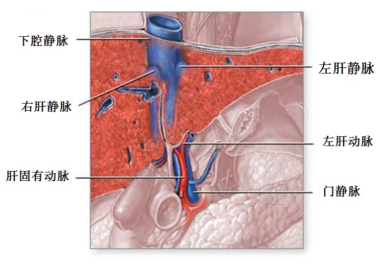
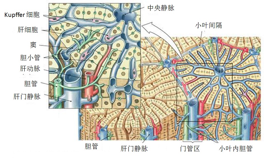
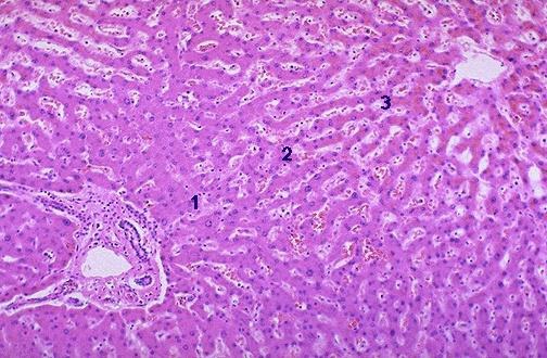
单支主肝静脉阻塞临床上一般无症状【2】。两或三支主肝静脉阻塞导致两种血流动力学的改变:增加肝窦血压和减少肝窦血流。肝血窦压力增加通过几个机制解释临床上的肝肿大、疼痛和腹水:(1)主要在肝小叶中央区实质上持续的血窦扩张和充血【2,3】;(2)当淋巴引流超过其能力的时候导致其通过肝包膜间质液体滤过增加【4】。通常,由于肝血窦壁对蛋白的高渗透性,滤过液内有较高的蛋白含量;(3)门静脉压增加【5】。
Budd Chiari's Syndrom
 |
|
Hepatic venous outflow obstruction |
有关全肝血流的变化还没有得到很好的阐述。间接证据表明伴随流出道受阻区域的微循环,门静脉血流灌注下降,动脉血的灌注增加【6-8】。这些变化的分布是不均匀的。肝细胞缺血损伤(非炎性中央小叶细胞坏死)占约70%【2】。肝细胞损伤可以由再灌注损伤引起【9】。中央小叶坏死可能由于充血而加重【10】。当同一区域的肝静脉和门静脉都阻塞,融合细胞(confluent cell)的损失发生在由末稍前(preterminal)和较大管区供应的区域【11】。缺血性肝细胞坏死导致的肝衰竭罕有爆发性的。肝静脉阻塞后的几个星期内肝纤维化形成可以在小叶中心区(当纯粹肝静脉阻塞的时候)或汇管区(有相关的门静脉阻塞)【3,11】。几个月内,发生结节样增生(nodular regeneration)主要发生在汇管区【2,3,11】。最终导致肝硬化[2,11]。大再生结节在约40%的患者中得到发现,特别是在减少门静脉灌注的区域[11,12]。单纯肝静脉阻塞而没有下腔静脉血栓的病人中肝细胞肝癌少见【11】。长期情况下大再生结节的恶性变仍然是一个悬而未决的问题。
 |
|
肝静脉流出道梗阻→肝窦压力增加→肝灌注下降→充血性门静脉高压→肝功能下降+细胞坏死+纤维化+肝硬化+结节性再生性增殖(nodular regeneration dysplasia) |
BCS病人中10%~20%伴有门静脉主干的阻塞【13】。肝移植时切除肝一半以上的标本可以发现肝内门静脉严重阻塞,主要是由于血流停滞和潜在的凝血倾向【11】。
由于肝静脉脉阻塞通常是不同步的,早期受累肝脏区域的萎缩与肝充血或后期受累肝脏区域的增生性肥大可以是共存的。一半的病例尾叶是肥大的,并可以引起下腔静脉狭窄【14,15】。尾叶的肥大主要由于其静脉引流是直接进入下腔静脉,同时也是阻塞的主肝静脉侧支循环的通道。
虽然有证据表明肝血窦压力增加是稳定和持久的状态,肝灌注减少似乎是断断续续(其判断来自于小叶中央坏死)和暂短的(其判断来自于血清转氨酶)。能够代偿灌注减少的自然机制可能参与其中,包括(1)动脉血流的增加;(2)门静脉压力的增加;(3)门静脉血流的重新分配(流出道损伤的区域朝向流出道保留的区域);(4)绕过阻塞的静脉形成侧支循环通道【16,17】。
-
Akiyoshi H, Terada T. Centrilobular and perisinusoidal fibrosis in experimental congestive liver in the rat. J Hepatol 1999; 30:433-439
-
Witte CL, Witte MH, Dumont AE. Lymph imbalance in the genesis and perpetuation of the ascites syndrome in hepatic cirrhosis. Gastroenterology 1980;78:1059-1068
-
Beattie C, Sitzmann JV, Cameron JL. Mesoatrial shunt hemodynamics. Surgery 1988;104:1-9
-
Menu Y, Sebag G, Vilgrain V, et al. Budd-Chiari syndrome: MR evaluation. Diagn Int Radiol 1990;2:23-28
-
Miller WJ, Federle MP, Straub WH, et al. Budd-Chiari syndrome: imaging with pathologic correlation. Abdom Imaging 1993;18:329-335
-
Soyer P, Rabenandrasana A, Barge J, et al. MRI of Budd-Chiari syndrome. Abdom Imaging 1994;19:325-329
-
Gonzalez-Flecha B, Reides C, Cutrin JC, et al. Oxidative stress produced by suprahepatic occlusion and reperfusion. Hepatology 1993;18:881-889
-
Henrion J. Ischemia/reperfusion injury of the liver: pathophysiologic hypotheses and potential relevance to human hypoxic hepatitis. Acta Gastroenterol Belg 2000;63: 336-347
-
Tanaka M, Wanless IR. Pathology of the liver in Budd-Chiari syndrome: portal vein thrombosis and the histogenesis of veno-centric cirrhosis, veno-portal cirrhosis, and large regenerative nodules. Hepatology 1998;27:488-496
-
Vilgrain V, Lewin M, Vons C, et al. Hepatic nodules in Budd-Chiari syndrome: imaging features. Radiology 1999; 210:443-450
-
Mahmoud AE, Helmy AS, Billingham L, et al. Poor prognosis and limited therapeutic options in patients with Budd-Chiari syndrome and portal venous system thrombosis. Eur J Gastroenterol Hepatol 1997;9:485-489
-
Gupta S, Barter S, Phillips GW, et al. Comparison of ultrasonography, computed tomography and 99mTc liver scan in diagnosis of Budd-Chiari syndrome. Gut 1987;28:242-247
-
Tavill AS, Wood EJ, Kreel L, et al. The Budd-Chiari syndrome: correlation between hepatic scintigraphy and the clinical, radiological, and pathological findings in nineteen cases of hepatic venous outflow obstruction. Gastroenterology 1975;68:509-518
-
Cho KJ, Geisinger KR, Shields JJ, Forrest ME. Collateral channels and histopathology in hepatic vein occlusion. AJR 1982;139:703-709
-
Hadengue A, Poliquin M, Vilgrain V, et al. The changing scene of hepatic vein thrombosis: recognition of asymptomatic cases. Gastroenterology 1994;106:1042-1047
-
Hirshberg B, Shouval D, Fibach E, et al. Flow cytometric analysis of autonomous growth of erythroid precursors in liquid culture detects occult polycythemia vera in the Budd-Chiari syndrome. J Hepatol 2000;32:574-578
|


