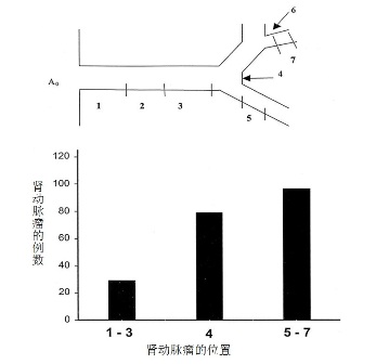
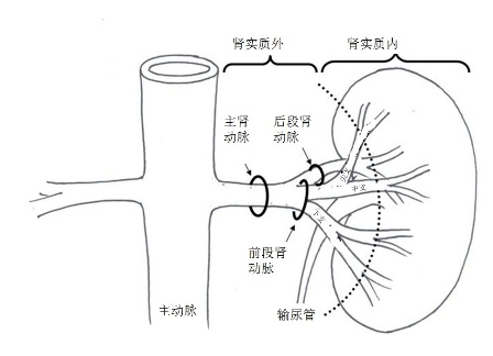
肾动脉直径的测量取决于所使用的影像工具。一项研究显示超声测量肾动脉平均直径为5.04±0.74mm;血管造影为5.68±1.19mm。这项研究中副肾动脉出现时,主肾动脉的直径明显小于只有一支肾动脉的时候[31]。 肾动脉瘤被定义为肾动脉段扩张超出受累动脉直径的两倍。其可以分为肾实质外动脉瘤和肾实质内动脉瘤(Intraparenchymal renal artery aneurysms ,IPRAAs)。IPRAAs可以同时存在狭窄性病变和实质外肾动脉瘤(extraparenchymal aneurysms of the renal artery)和肾癌[1~6]。实质外肾动脉瘤占有全部肾动脉瘤的大部分(85%)[1,27]。其余为肾实质内动脉瘤约为15%。实质外肾动脉瘤70%为囊性,20%为梭形,10%为夹层[32]。 肾动脉瘤病人,20%表现为双侧肾动脉瘤,而30%是多发动脉瘤[1]。男女发病率相等,虽然育龄女性的肾动脉瘤更容易破裂。 肾动脉瘤的自然病史并不十分清楚,但有潜在的并发症包括动脉瘤破裂和肾栓塞[27]。 破裂的危险因素包括不完全钙化,直径大于2cm,进行性增大和妊娠。
肾动脉瘤相关解剖
(1)真性动脉瘤
Ehlers-Danlos,是常染色体显性遗传性疾病,特点是动脉中层弱化,主要由于缺乏III型前胶原。这种状况导致包括肾动脉的任何动脉发生动脉瘤和/或动脉夹层[21]。 在假性动脉瘤,是局限性的动脉壁一层或全层中断,在被弱化的部位导致动脉囊状外凸。在顿性损伤时,相对活动的肾脏向前移位,在血管蒂产生张力形成内膜撕裂,造成内膜下夹层或动脉瘤形成。或者椎体直接损伤动脉壁[22]。 来自于以前肾动脉重建手术的吻合口漏可以导致假性动脉瘤。这种情况下,动脉瘤壁仅包含纤维/炎性组织。医源性血管内治疗有关的动脉瘤是由于内膜创伤和局部夹层,导致动脉瘤样变性。自发性肾动脉夹层也可以引起动脉瘤[23]。 实质内动脉瘤被认为是主要的血管壁产生的炎性改变,通常表现为小的动脉瘤。 虽然怀孕与肾动脉瘤发病率增加没有相关性,但怀孕与肾动脉瘤破裂的关联率较高[26]。增加的血流量,腹内压力和由于妊娠有关的激素及代谢变化导致血管壁的弱化被认为是主要原因。大多数破裂发生在妊娠晚期,妊娠末三个月,并左肾动脉较多[9]。
在儿童,肾动脉瘤是由于外伤,感染,动脉炎,川崎病,或血管发育不良。多动脉受累的多发性、特发性肾动脉动脉瘤已被描述,但非常罕见[26]
肾实质动脉瘤  肾动脉瘤在每个肾动脉段的位置。多数肾动脉瘤位于肾实质内的动脉分叉位置[1] 感染性肾动脉瘤:并不常见。发生率依次为主动脉、周围血管、脑血管和内脏的动脉。葡萄球菌和链球菌最常见的病原菌,还有真菌感染。真菌性动脉瘤是由于微生物进入血循环导致血管壁坏死的结果,通常是细菌性心内膜炎[9]。确切的发病率不清楚,可能是细菌性心内膜炎病人的1~10%。而金黄色葡萄球菌或链球菌感染性动脉瘤在20~36%的病人[7]。一般来说,感染性动脉瘤由于出血和败血症都有一个致命的自然病史。
2. Hageman JH, Smith RF, Szilaghi DE, Elliott JP. Aneurysm of the renal artery: problems of prognosis and surgical management. Surgery 1978; 84: 563–572. 3. Henke PK, Cardneau JD, Welling TH et al. Renal artery aneurysm. A 35-year clinical experience with 252 aneurysms in 168 patients. Ann Surg 2001; 4: 454–463. 4. Barett JM, Dean RH, Boehm FH. Rupture of renal artery aneurysm during pregnancy. South Med J 1981; 74: 1549. 5. De Bakey ME, Lefrak EA, Garcia-Rinaldi R, Noon GP. Aneurysm of the renal artery. A vascular reconstructive approach. Arch Surg 1973; 106: 438–443. 6. Hafez KS, Krishnamurthi V, Campbell SC, Novick AC. Contemporary management of renal cell carcinoma with coexistent renal artery disease: update of the Cleveland Clinic experience. Urology 2000; 56: 382–386. 7. Henkes H, Terstegge K, Felber S, Jänisch W, Nahser HC, Kühne D (1993) Mykotisches, infektionsbedingtes intrakranielles aneurysma. In: Henkes H, Kölmel HW (eds) Die entzündlichen erkrankungen des zentralnervensystems. Ecomed, Landsberg, Germany, pp 1–71 8. Cerny JC, Chang CY, Fry WJ. Renal artery aneurysms. Arch Surg 1968; 96: 653–663. 9. Cohen JR, Shamash FS. Ruptured renal artery aneurysms during pregnancy. J Vasc Surg. Jul 1987;6(1):51-9. 10. Pautasse EF. Renal artery aneurysms. J Urol 1976; 113: 443–449. 11. Mercier C, Piquet P, Piligian F, Ferdan M. Aneurysm of the renal artery and its branches. Ann Vasc Surgery 1986; 1: 321–327. 12. Chen R, Novick AC. Retroperitoneal hemorrhage from a rupterd renal artery aneurysm with spontaneous resolution. J Urol 1994; 151: 139–141. 13. Smith JN, Hinman F Jr. Intrarenal arterial aneurysms. J Urol 1967; 97: 990–996. 14. Hageman JH, Smith RF, Szilaghi DE, Elliott JP. Aneurysm of the renal artery: problems of prognosis and surgical management. Surgery 1978; 84: 563–572. 15. Tham G, Ekelund L, Herrlin K, Lindstedt EL, Olin T, Bergenntz SE. Renal artery aneurysm. Natural history and prognosis. Ann Surg 1983; 197: 348–352. 16. Goldfarb M, Kase S. Renal microaneurysms and hematuria. Urology 1975; 6: 86–88. 17. Longstreth PL, Korobkin M. Intrarenal arterial aneurysms. Crit Rev Clin Radiol Nucl Med 1976; 8: 129–151. 18. Ortenberg J, Novick AC, Straffon RA, Stewart Bh. Surgical treatment of renal artey aneurysms. Br J Urol 1983; 55: 341. 19. Vaugham TJ, Barry WF, Jeffords DL, Johnsrude IS. Renal artey aneurysm and hypertension. Radiology 1971; 99: 287–293. 20. McCarron JP, Marshall VF, Whitsell JC. Indications for surgery of renal artery aneurysms. J Urol 1975; 114: 177–180 21. Mattar SG, Kumar AG, Lumsden AB. Vascular complications in Ehlers-Danlos syndrome. Am Surg. Nov 1994;60(11):827-31. 22. Gewertz BL, Stanley JC, Fry WJ. Renal artery dissections. Arch Surg. Apr 1977;112(4):409-14. 23. Calligaro KD, Dougherty MJ. Renal artery aneurysms and arteriovenous fistulae. In: Rutherford RB, ed. Vascular Surgery. 5th ed. Philadelphia, Pa: WB Saunders; 2000:1697-702. 24. Callicutt CS, Rush B, Eubanks T, et al. Idiopathic renal artery and infrarenal aortic aneurysms in a 6-year-old child: case report and literature review. J Vasc Surg. May 2005;41(5):893-6 27. Lumsden AB, Salam TA, Walton KG: Renal artery aneurysm: a report of 28 cases. Cardiovasc Surg 1996 , 4(2):185-189 28. Seppala F, Levey J: Renal artery aneurysm: case report of a ruptured calcified renal artery aneurysm. Am J Surg 1982 , 48:42-44 29. Lacroix H, Bernaerts P, Nevelsteen A, Hanssens M: Ruptured renal artery aneurysm during pregnancy: successful ex situ repair and autotransplantation.J Vasc Surg 2001 , 33(1):188-190. 30. Stephens, FD. Smith, ED, Hutson, JM. Ureterovascular hydronephrosis and the aberrant renal vessels. In: Congenital Anomalies of the Kidney, Urinary and Genital Tracts. 2nd. New York, New York: Informa Health Care; 2002:275-80. 31. Aytac SK, Yigit H, Sancak T, et al. Correlation between the diameter of the main renal artery and the presence of an accessory renal artery: sonographic and angiographic evaluation. J Ultrasound Med. May 2003;22(5):433-9; quiz 440-2. 32. Bastounis E, Pikoulis E, Georgopoulos S, et al. Surgery for renal artery aneurysms: a combined series of two large centers. Eur Urol. 1998;33(1):22-7 33. Henriksson C, Bjorkerud S, Nilson AE, et al. Natural history of renal artery aneurysm elucidated by repeated angiography and pathoanatomical studies. Eur Urol. 1985;11(4):244-8.
|


